Raman spectroscopy and microscopy

Raman spectroscopy and microscopy
© Fraunhofer IGB
Raman spectroscopy and microscopy.
© Fraunhofer IGB
Raman spectroscopy and microscopy.

Confocal Raman microspectroscopy enables spatially resolved identification of chemical, biological and mineral components in complex heterogeneous samples.


Measurement principle and areas of application

Similar to infrared spectroscopy, Raman spectroscopy uses the interaction of light with matter, i.e. the capture of molecular vibration bands on irradiation with monochromatic light coming from a laser, which allows insights into the molecular structure and properties of a material. A major advantage of Raman spectroscopy is the application in aqueous media due to no interfering water band in the spectrum.

In principle, Raman-based analysis and characterization can be applied to a wide variety of materials, from polymers as well as medical and pharmaceutical products to biological tissue samples.


Automated and highly flexible Raman analyses with Leica microscopes and CARS unit

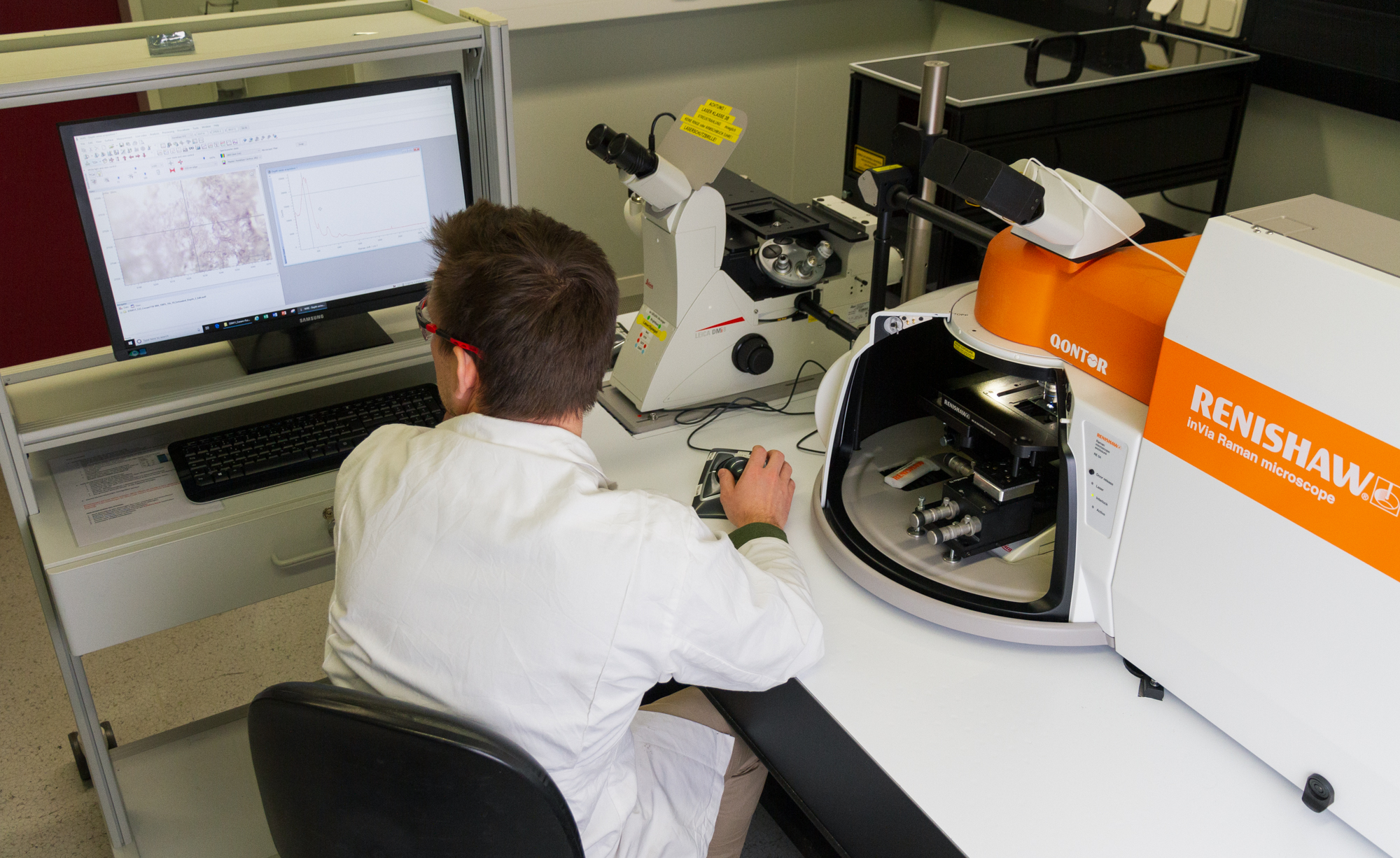
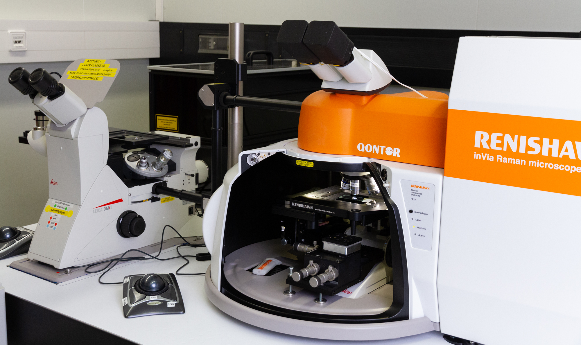
With the confocal inVia™ Qontor Raman microscope from Renishaw, Fraunhofer IGB has the opportunity to use an automated and highly flexible microscope for research which can be used for investigations in all business areas of the IGB: health, environment and sustainable chemistry.

The system is equipped with two Leica microscopes that allow measurements in the upright position (with chamber) and inverse in transmission. For maximal flexibility, the system can use a 532 nm (max. 50 mW) and 785 nm (max. 300 mW) laser as well as a CARS unit (coherent anti-Stokes Raman spectroscopy). The CARS unit offers the opportunity to filter unwanted fluorescent signals resulting from the sample. Being one of the first, Fraunhofer IGB can perform measurements with a laser and CARS-unit almost simultaneously on the same sample.


Analysis of chemical compounds and materials

The most common application of Raman microscopy is the analysis of chemical compounds (e.g. materials, catalysts, liquids, etc.) In the past, the IGB looked at different hydrogels.


Identification of substances in samples of unknown composition

Additionally, band behavior from Raman microscopy allows the non-destructive identification of the chemical composition of an unknown sample. Here, Fraunhofer IGB was able to determine the formation mechanism of chemical corrosion in a copper sample and thus was able to exclude a biological reason for material destruction.


Qualitative and quantitative analysis of cells in biological samples

In the field of biological research, Raman spectroscopy is gaining increased significance as well. The main field of application of the technology is the analysis of cell systems because the analysis can be performed without sample preparation and is non-destructive. Current research at IGB includes differentiation of microorganisms in biofilms, differentiation of live and dead cells as well as quantification of microorganisms in fluidic systems.

Application examples

Identification of contaminations on a microchip

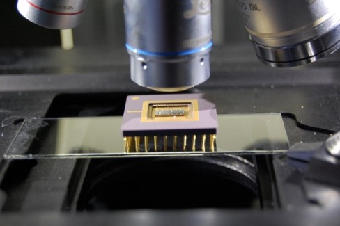
Examination of a microchip, the sample site is located in a depression of approx. 3 mm.
© Fraunhofer IGB

Examination of a contaminated microchip, the sample site is located in a depression of approx. 3 mm.

Using a 50x objective with 8.5 mm LWD (long working distance), suitable measuring points could be identified and spatially resolved Raman spectroscopic investigations performed.

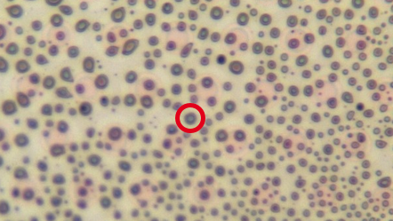
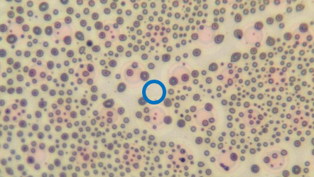
Contaminated measuring point (red).
© Fraunhofer IGB
Contaminated measuring point (red).
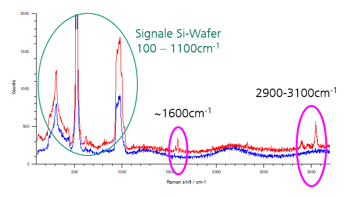
Raman spectra of the contaminated (red) and uncontaminated (blue) measurement sites with 532 nm laser. The marked bands at 1600 cm<sup>-1</sup> and 3050 cm<sup>-1</sup> indicate aromatic components of the approx. 1 µm droplet-shaped contaminants.
© Fraunhofer IGB
Raman spectra of the contaminated (red) and uncontaminated (blue) measurement sites with 532 nm laser. The marked bands at 1600 cm-1 and 3050 cm-1 indicate aromatic components of the approx. 1 µm droplet-shaped contaminants.
Not contaminated measuring point (blue).
© Fraunhofer IGB
Not contaminated measuring point (blue).

Raman spectroscopy for microbiological analysis

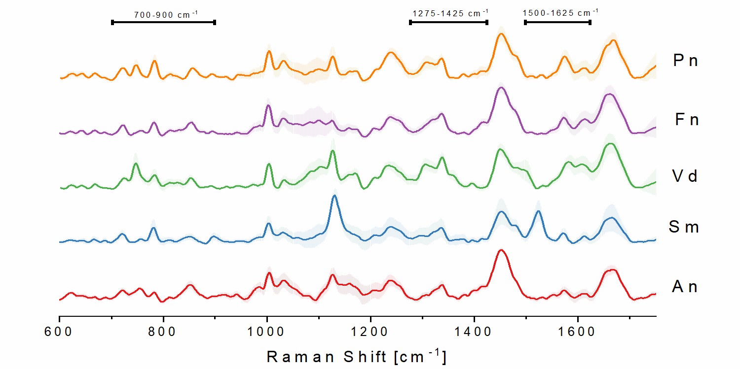
Differentiation of oral bacteria using Raman spectra and statistical models.
© Fraunhofer IGB
Differentiation of oral bacteria using Raman spectra and statistical models.

Different oral bacterial strains were cultured under the same conditions, fixed on glass coupons and measured with a 532 nm laser. Spectra were processed to visualize specific bands in the 600–1800 nm-1 region ("fingerprint" region for organic samples where chemical differences are visible).

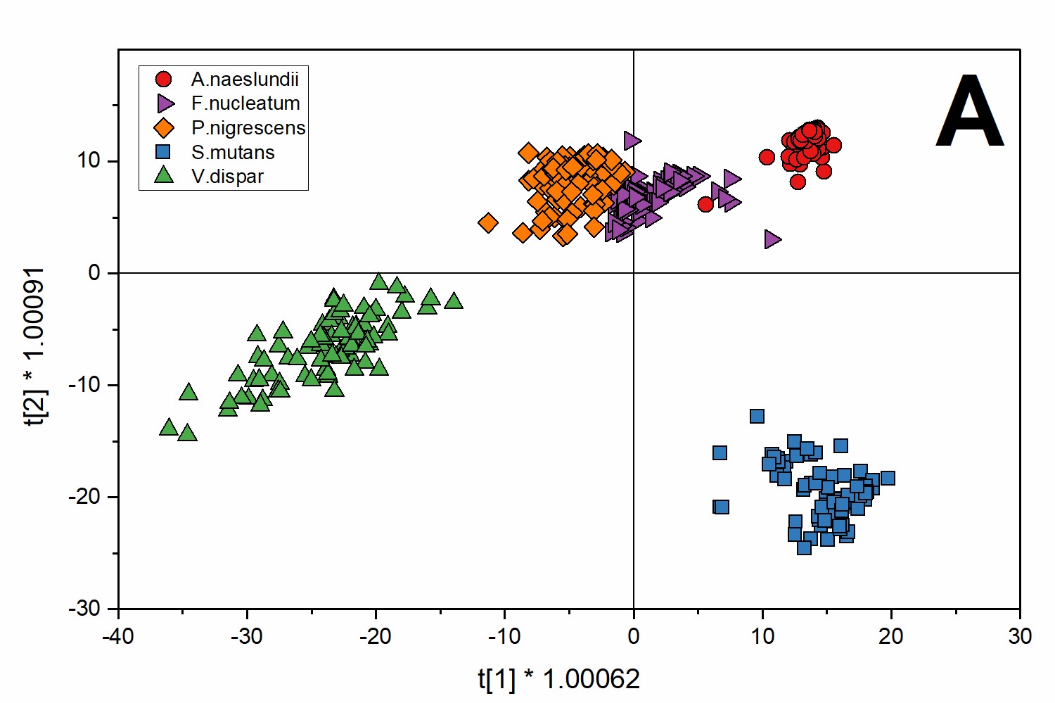
Using a statistical model (in this case O2PLS-DA) based on the collected spectra, differences between the bacteria were detected. These differences are due to different banding behavior in the "fingerprint" region.
© Fraunhofer IGB

Using a statistical model (in this case O2PLS-DA) based on the collected spectra, differences between the bacteria were detected. These differences are due to different banding behavior in the "fingerprint" region.

Determining causes of corrosion

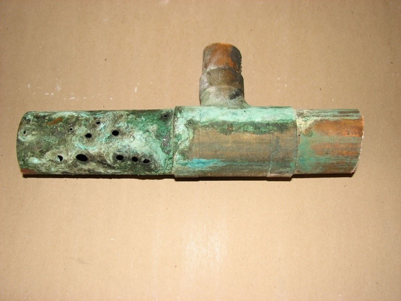
Example of corrosion on a pipe (does not correspond to the actual sample).
© Fraunhofer IGB
Example of corrosion on a pipe (does not correspond to the actual sample).

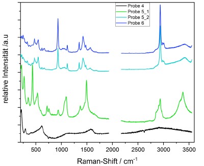
Three differently corroded pipe samples showing different visual characteristics were examined. The aim was to determine what type of corrosion was present. The pipe samples were collected from a production plant.

Raman-Analyse gekoppelt mit FTIR-Analyse
© Fraunhofer IGB

Raman analysis coupled with FT-IR analyses allowed a clear classification of the corrosions. Copper oxide (sample 4) and malachite (samples 5 and 6) can be seen. Based on the Raman spectra, additional impurities of copper acetate and copper formate were detected.

Publications

Kriem, L.S.; Wright. K.; Ccahuana-Vasquez, R.A.; Rupp, S. (2021) Mapping of a Subgingival Dual-Species Biofilm Model Using Confocal Raman Microscopy, Front. Microbiol. 12:729720. doi: 10.3389/fmicb.2021.729720

Short summary: Microorganisms tend to attach to surfaces, multiply and form biofilms. Oral biofilms have previously been studied in the context of various oral diseases. In a new publication, Lukas Kriem and others describe an alternative method to visualize the architecture of oral biofilms that form under the gums. This can help to better understand disease mechanisms.

Kriem, L.S.; Wright. K.; Ccahuana-Vasquez, R.A.; Rupp, S. (2020)
Confocal Raman microscopy to identify bacteria in oral subgingival biofilm models, PLoS ONE 15(5): e0232912. https://doi.org/10.1371/journal.pone.0232912