Bioprinting – Optimized bioinks for additive manufacturing

Medicine of the future is biological. In order to turn the concept of biological implants and personalized therapies into tangible reality, scientists at Fraunhofer IGB are optimizing biological materials for processing with 3D printing techniques.

Like classical tissue engineering, the so-called bioprinting pursues the goal of producing biological or biologically functional tissue in the laboratory. The printed in vitro tissues will serve as test systems to answer questions about the efficacy of active substance candidates and thus help replace animal testing. In the future, as biological implants, they may also act to stimulate damaged tissue to regenerate, or substitute them. 

Physiological tissues from printed biomaterials

“We must create an environment where body cells isolated from tissues and multiplied in the laboratory can fulfill their specific functions over a longer period of time,” says Dr. Kirsten Borchers about one of the challenges facing the scientists. The best artificial environment for the cells is one that mimics the natural conditions in the body as closely as possible: “In our printed tissues, the role of the tissue matrix is thus taken over by biomaterials that we generate from molecules of the natural tissue matrix,” explains the scientist.

In order for the biomaterials to be brought into shape by means of 3D printing, their flow properties must be adapted to the printing technique used in each case. After the printing process, the structure thus generated is then additionally stabilized by a cell-compatible crosslinking reaction. Dr. Borchers and her colleagues, Prof. Dr. Günter Tovar and Dr. Achim Weber and her team, set out to meet these challenges.

Flexible shapes and standardized conditions through additive manufacturing

"In order to build tissues in the lab that work just as well as their natural counterparts, we need not only the best possible biomaterials, but also production processes that limit us as little as possible in terms of shaping," explains Borchers.

“Additive methods offer great flexibility in this respect. With their help, three-dimensional objects that were previously designed on the computer can now be fabricated layer by layer,” says Weber. Flexible shaping is particularly important in terms of the vision of generating personalized printed biological implants. Because modern diagnostic imaging modalities, which are currently already frequently used in daily medical practice, can provide the very digital 3D data required for customized implants.

“The additive processing of tissue matrix and cells into tissue models offers yet another advantage – automation and digital control could in the future guarantee that production can take place under controlled and standardized conditions,” adds Borchers.

In the current state of the art, 3D printing methods are used to combine different cells and biomaterials as well as structures such as perfusion channels for suppling the cells in simply constructed tissue models [1]. The branch of research that deals with the fabrication of biological structures using additive manufacturing processes is known as bioprinting.

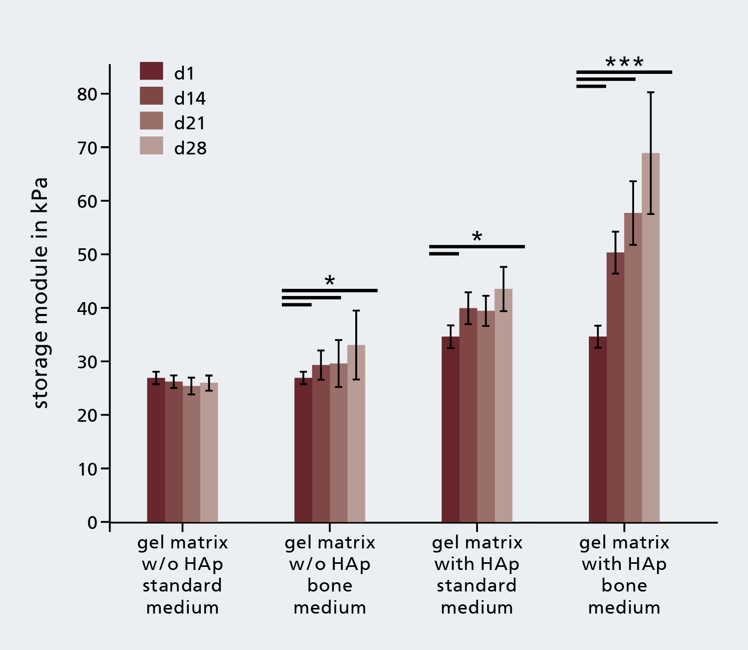
The addition of hydroxylapatite (HAp) to bone ink increases remodeling of the matrix during the culture of osteogenically diferentiated mesenchymal stem cells.
© Fraunhofer IGB
The addition of hydroxylapatite (HAp) to bone ink increases remodeling of the matrix during the culture of osteogenically diferentiated mesenchymal stem cells.

A material kit with natural biopolymers

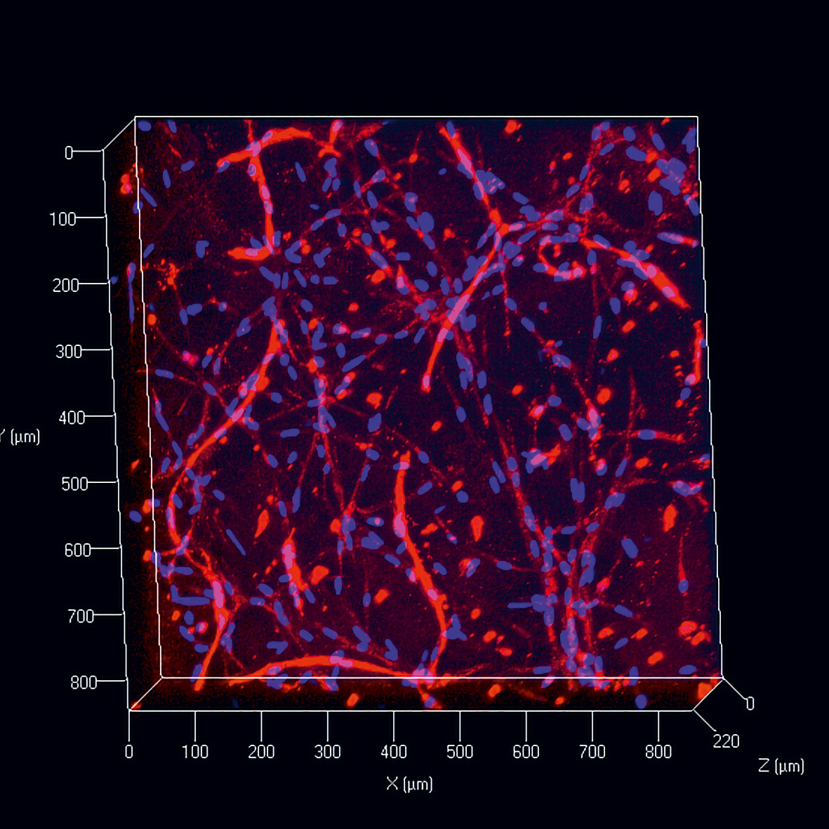
Capillary formation in a vascularization ink.
© Fraunhofer IGB
Capillary formation in a vascularization ink.

Typically, the tissue matrix has the texture of a highly hydrous gel. In bones, mineral components are embedded as well. In this way, the extracellular matrix (ECM) ensures the mechanical stability of tissues. In addition, the tissue matrix is also involved in intercellular communication. It is not easy to simulate these comprehensive functions with synthetic materials.    

“Our approach is therefore to optimize biopolymers recruited from the natural portfolio for technical processing,“ says Borchers. Natural, biofunctional molecules of the tissue matrix such as gelatin, heparin, hyaluronic acid and chondroitin sulfate are chemically furnished with additional functions at IGB: “As an example, by ´masking´ certain side chains of biomolecules, we can reduce intermolecular interactions and thereby influence the viscosity and gelling behavior of gelatin solutions,“ explains the scientist. On the other hand, reactive groups can be introduced in order to chemically crosslink biomolecules to generate hydrogels, for example by means of a light stimulus. The ratio of the introduced masking acetyl and the reactive methacrylic groups enables both the flow behavior of the solutions and the swelling properties of the crosslinked hydrogels to be adjusted [2-3].

Optimized bioinks for biologically functional tissues

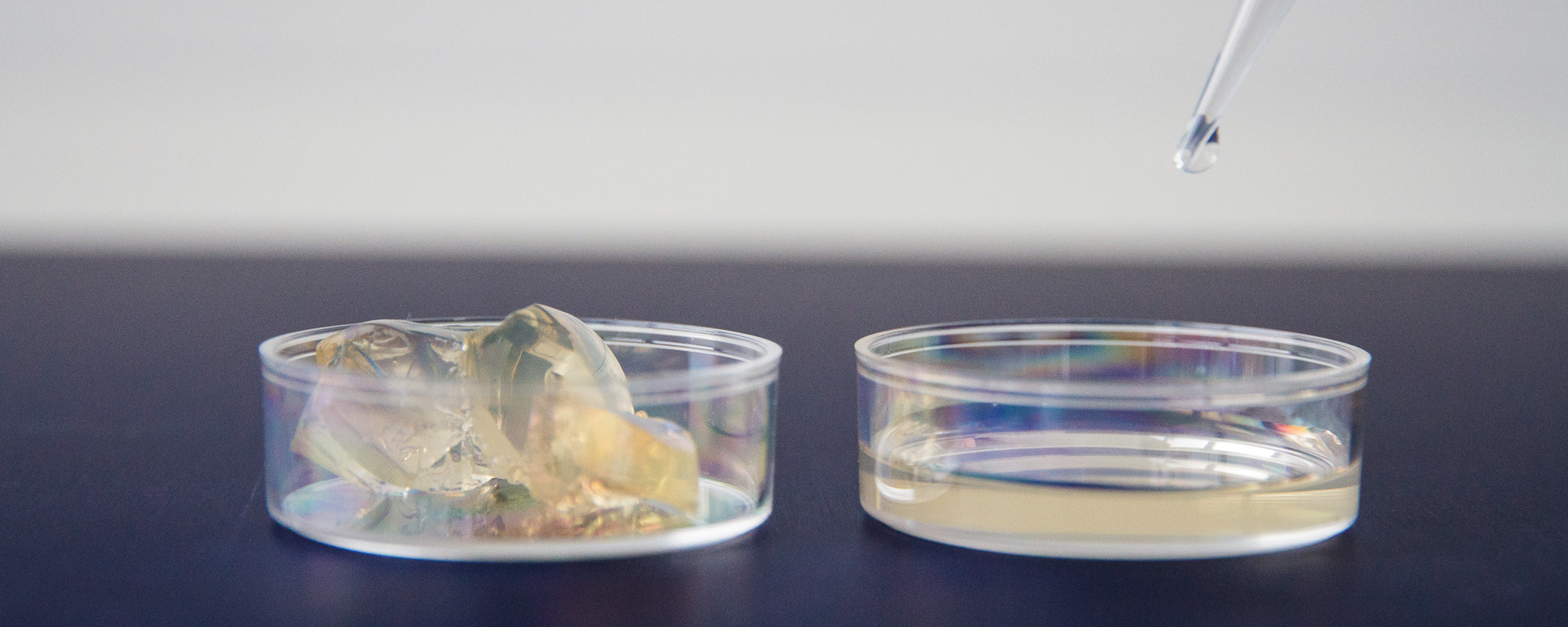
“Bioink“ is a biomaterial in its uncrosslinked, printable form. By deliberately varying the composition, bioinks are optimized for the printing process and at the same time for the stimulation of tissue-specific functions. Borchers and her colleagues were already able to successfully produce “bone inks“ and “vascularization inks“ on the basis of the available material kit. Both bioinks are dispersions of biomolecules and tissue-typical cells that can be stably converted into a 3D structure via dispensing processes.

Bioinks for functional bones and vessels

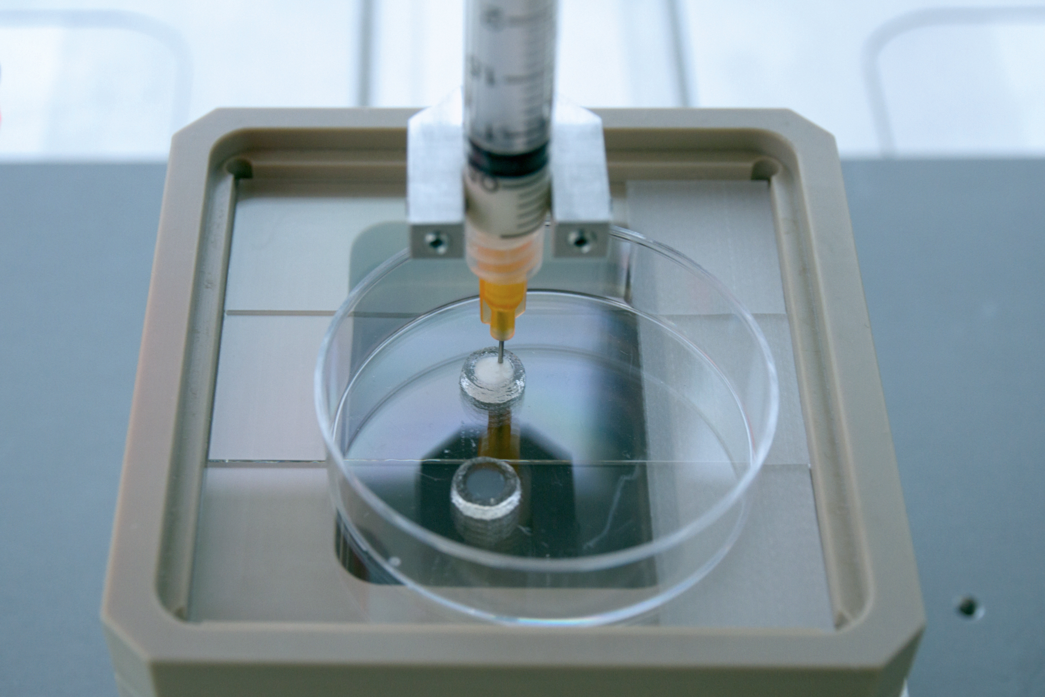
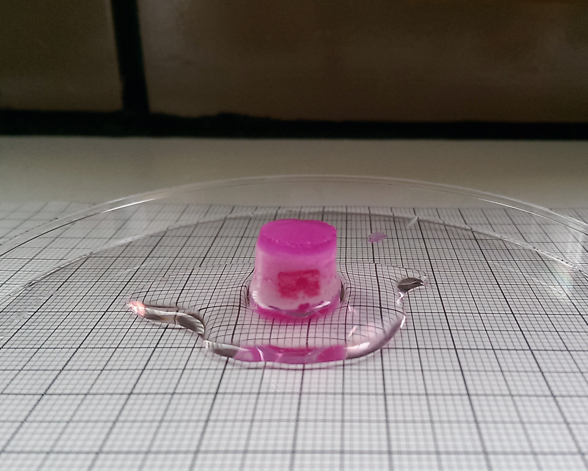
Individually or in combination, our bioinks can be used to construct vascularized tissue models.


Bone ink    

Bone ink contains a mass fraction of 13 percent of crosslinkable biopolymers and a mass fraction of 5 percent hydroxylapatite (HAp) as a tissue-specific mineral additive. The proportion of HAp is adjusted in such a way that the vitality of the mesenchymal stem cells used and the crosslinking reaction of the hydrogels are not impaired [4]. The increase in the viscosity of the ink due to the addition of the HAp is very desirable: By choosing a suitable ratio of the available gelatin derivatives with different gelling abilities, a gelling temperature of, say, 21.5°C can then be set. Bone ink thus has excellent extrudability at room temperature.

Research has shown that after the ink has been crosslinked to hydrogel, the mineral component promoted the remodeling of the matrix by the cells contained therein: The mechanical strength of the gels increased more markedly during the four-week cultivation when HAp was contained in the matrix than in carrier gels without HAp [5]. Raman spectroscopy suggests that the observed effect is mainly due to increasing mineralization of the matrix. In addition, bone-typical marker proteins indicate that the mesenchymal stem cells differentiated into bone cells in the printed matrices. 

Vascularization ink

The supply of nutrients and oxygen via vascular structures is particularly important for extensive in vitro tissues, as diffusion takes too long. The endothelial cells that line the vessels from the inside play an important role in the formation and growth of new vessels.

Vascularization matrix must have different properties than bone matrix: First and foremost, it must be soft and less strongly cross-linked, so that the endothelial cells can migrate and form capillaries. The vascularization ink developed at IGB therefore contains only 5.75 percent by weight of crosslinkable biopolymers. These also have a low degree of methacrylate and thus crosslink less strongly than bone ink. By the addition of gelatin derivatives with masking, IGB succeeded in manufacturing soft vascularization gels (storage modulus of 2.7 kPa ± 0.31 kPa) with a high water absorption capacity (degree of swelling in equilibrium > 2000 percent). By varying the share of unmodified gelatin (which gels already at room temperature), it was possible to produce a bioink that can be stably printed at room temperature. When microvascular endothelial cells are introduced into these gels, the formation of capillary-like structures takes place.

Tissue model for vascularized bone.
© Fraunhofer IGB
Tissue model for vascularized bone.

Literature

  1. Kang, H.-W.; Lee, S. J.; Ko, I. K.; Kengla, C.; Yoo, J. J.; Atala, A. ( 2016) A 3D bioprinting system to produce human-scale tissue constructs with structural integrity, Nature Biotechnology 34: 312-319.
  2. DE 10 2012 219 691 B4 Modifizierte Gelatine, Verfahren zu ihrer Herstellung und Verwendung (2014).
  3. Hoch, E.; Hirth, T.; Tovar, G.; Borchers, K. (2013) Chemical tailoring of gelatin to adjust its chemical and physical properties for functional bioprinting, Journal of Materials Chemistry B 1: 5675-5685.
  4. Wenz, A.; Janke, K.; Hoch, E.; Tovar, G.; Borchers, K.; Kluger, P. (2016) Hydroxylapatite-modified gelatin bioinks for bone bioprinting, BioNanoMaterials 17: 179-184.
  5. Wenz, A.; Borchers, K.; Tovar, G.; Kluger, P. (2017) Bone matrix production in hydroxylapatite-modified hydrogels suitable for bone bioprinting, Biofabrication 9: 044103.    

Research field of bioprinting

For several years, Fraunhofer IGB has been addressing bioprinting. In the Fraunhofer project BioRap, the foundations were first laid for additive manufacturing with biological cells and biomaterials that were already used in the EU project ArtiVaSc.    

Collaboration with University of Stuttgart   

This scientifically demanding and challenging field of research implicitly requires the investigation of fundamental issues. IGB therefore works closely with its partner institute at the University of Stuttgart, the Institute of Interfacial Process Engineering and Plasma Technology (IGVP).

Here, Dr. Kirsten Borchers, along with Prof. Dr. Günter Tovar (and until 2017 also with Prof. Dr. Petra Kluger), supervise doctorate students and student graduation theses in the field of biomaterial development. The presented results are part of Annika Wenz's PhD thesis (scholarship by Carl Zeiss Foundation) and Julia Rogal's Master's thesis.

We thank all donors for supporting our projects.