Three-Dimensional (3D) Skin Models as In-vitro Test Systems

We develop metabolically active 3D skin models of varying complexity as in vitro test systems based on immortalized primary or primary cells:

Reconstituted dermis models,

which are constructed from immortalized primary or primary fibroblasts in a collagen layer on a porous polycarbonate membrane and are cultivated submerged.

Reconstituted epidermis models,

which are constructed from immortalized primary or primary keratinocytes on a porous polycarbonate membrane and cultured at the air-liquid interface.

Reconstituted full-thickness skin models,

composed of immortalized primary or primary fibroblasts and immortalized primary or primary keratinocytes. The dermal tissue equivalent of fibroblasts is cultured on a porous polycarbonate membrane. This is covered by an epidermal layer of differentiated immortalized primary or primary keratinocytes. The model is cultivated at the air-liquid interface. 

Immunocompetent full-thickness skin models,

which are constructed from reconstituted full-thickness skin models, supplemented with immune cells such as peripheral blood mononuclear cells (PBMCs) and cultivated at the air-liquid interface on a porous polycarbonate membrane.

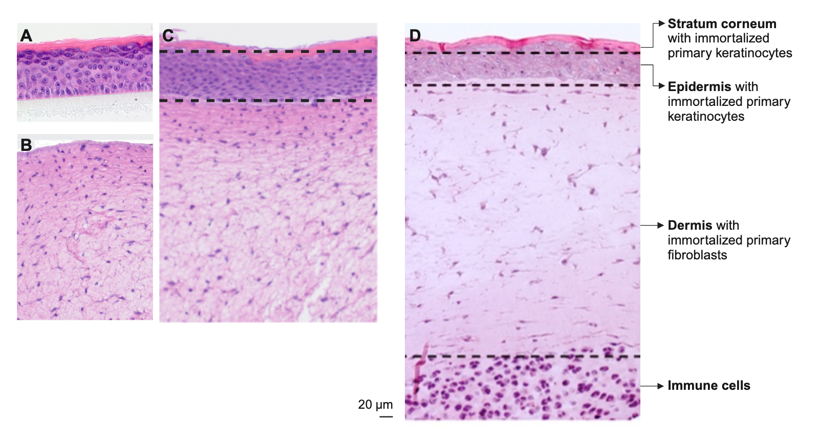
Histological sections of human 3D skin models: A) reconstituted epidermis model, B) reconstituted dermis model and C) reconstituted full-thickness skin model are tissue-typical. D) Immunocompetent skin model in which a full skin model was supplemented with immune cells in a collagen matrix.
© Fraunhofer IGB
Histological sections of human 3D skin models: A) reconstituted epidermis model, B) reconstituted dermis model and C) reconstituted full-thickness skin model are tissue-typical. D) Immunocompetent skin model in which a full skin model was supplemented with immune cells in a collagen matrix.

3D skin models of varying complexity from immortalized primary or primary cells

Challenge

Suitable alternatives to animal testing are so-called New Approach Methodologies (NAMs), which also include cell-based in-vitro models. 3D skin models are essential in the risk assessment of new products and in cosmetic research, where animal testing is completely banned in Europe.

Our development

With our decades of expertise in the development of monoclonal cell lines, the handling of (pathogenic) microorganisms and working to the highest quality standards, we develop 3D skin models of varying complexity, both for the representation of healthy skin and for its clinical pictures. Depending on the application, the skin models can contain immune cells, microorganisms and reporter systems for the activation of cellular signaling pathways in addition to skin components. 

This enables investigations into

  • on skin sensitization and toxicity, 
  • the immune response, 
  • host reactions of the skin in the presence of pathogenic microorganisms, 
  • interactions of the skin with the microbiome,
  • skin penetration and
  • mechanisms of action of substances.
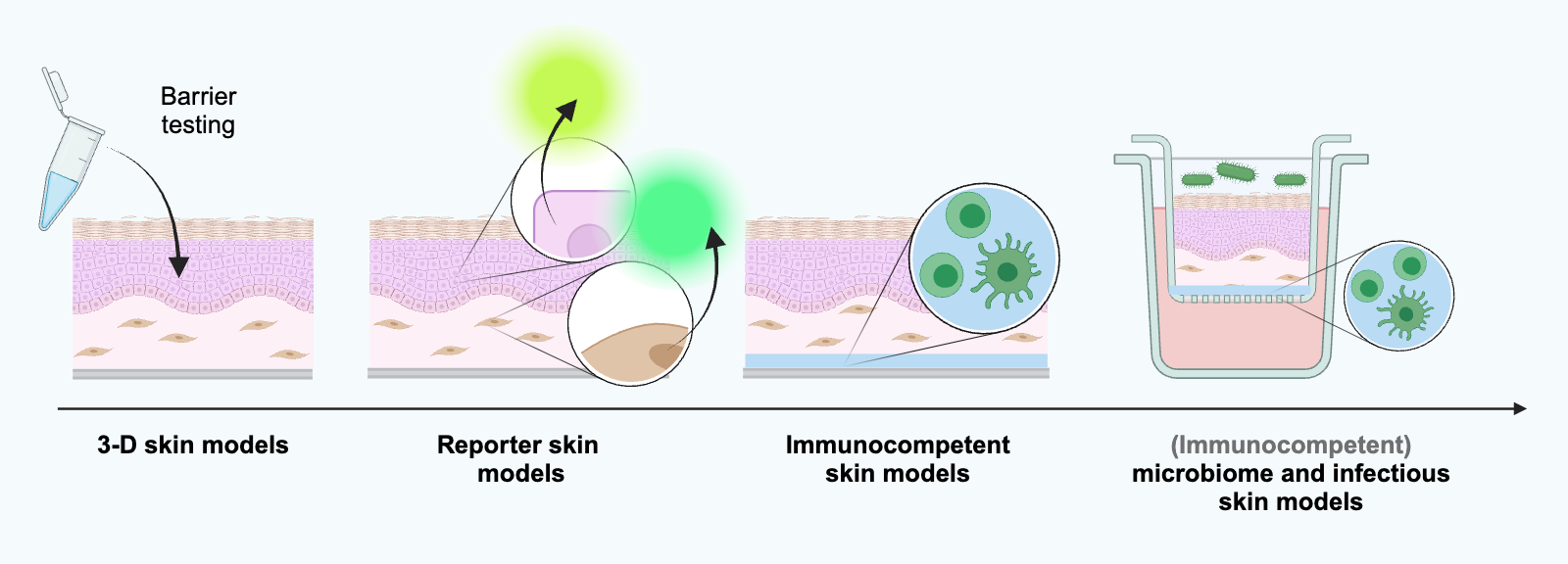
Schematic representation of 3D skin models of varying complexity as reporter skin models, immunocompetent models as well as microbiome and infection models (created with BioRender.com).
© Fraunhofer IGB
Schematic representation of 3D skin models of varying complexity as reporter skin models, immunocompetent models as well as microbiome and infection models (created with BioRender.com).

Applications of in vitro models

In dermatology and pharmacology, in-vivo studies on animals are still required before active ingredients and formulations can be tested on humans. However, new approach methodologies and in-vitro models in particular are becoming increasingly important here. Many results from animal studies, especially when the immune system is involved, are not directly transferable to humans. (Marshall LJ, Bailey J, Cassotta M, Herrmann K, Pistollato F. Poor Translatability of Biomedical Research Using Animals — A Narrative Review. Alternatives to Laboratory Animals. 2023;51(2):102-135. doi:10.1177/02611929231157756


In-vitro models enable 

  • a higher throughput, 
  • better species-specific prediction of in-vivo efficacy,
  • a deeper understanding of mechanisms of action by studying specific cell populations and 
  • support the selection of the best lead candidates. 

The use of in vitro models can accelerate drug development in both human and veterinary pharmacy.

3D skin models correspond to the in-vivo situation

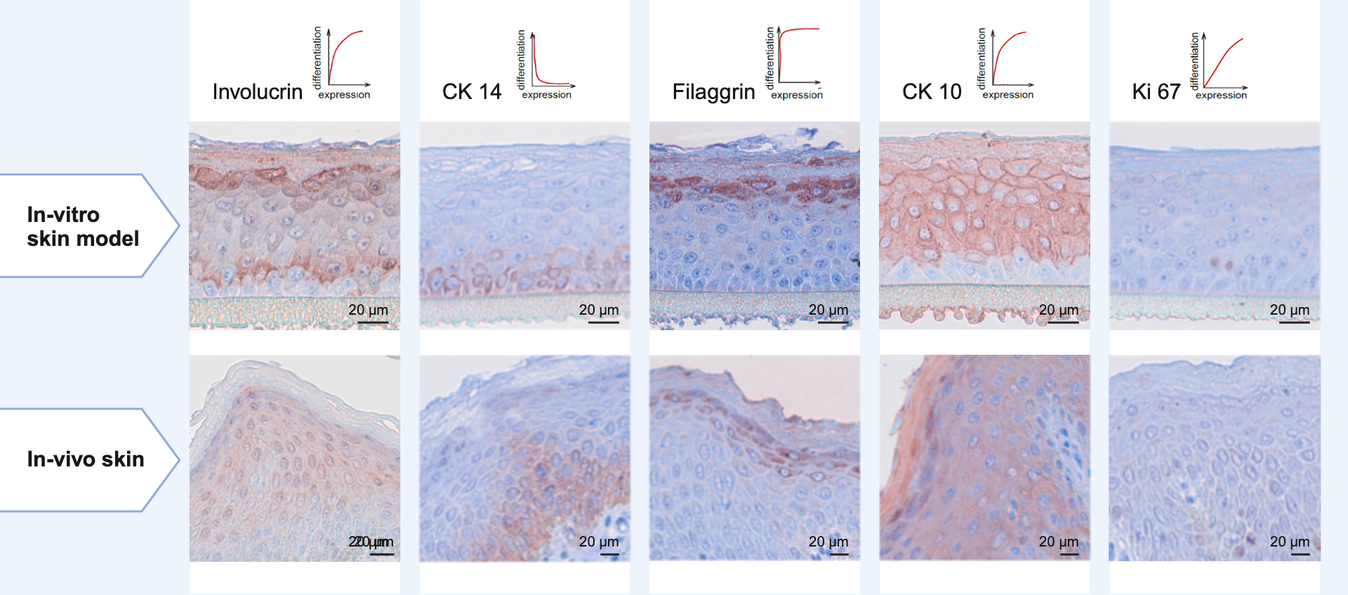
Our reconstituted epidermis and full-thickness skin models have a very pronounced horny layer with a barrier effect. Our models correspond to human in-vivo skin and express the proliferation marker Ki67 as well as specific differentiation markers of the epidermal layers exactly as in human skin:

  • Fillagrin
  • Involucrin
  • Cytokeratin 10
  • Cytokeratin 14
Proliferation and differentiation markers of reconstituted in vitro epidermis models correspond exactly to the expression pattern of human in vivo skin (immunohistochemical staining of tissue thin sections).
© Fraunhofer IGB
Proliferation and differentiation markers of reconstituted in vitro epidermis models correspond exactly to the expression pattern of human in vivo skin (immunohistochemical staining of tissue thin sections).

Advantages and unique selling points of our 3D skin models

Our 3D skin models are based on specifically immortalized primary or primary skin cells of human or animal origin from healthy donors. This makes our skin models donor-independent, reproducible and available for examinations at any time.

We establish models that carry a reporter function, mimic disease patterns or can be used to validate drug targets via knock-out. 

Our reconstituted epidermis models, which are cultivated without media additives of animal origin, enable batch-independent reproducibility. They are therefore a consistent solution for avoiding animal testing. 

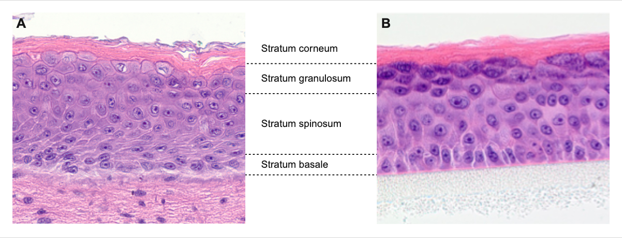
Histologische Schnitte A) eines rekonstituierten Vollhautmodells mit mehreren physiologischen Schichten bestehend aus Epidermis sowie Dermis und B) eines rekonstituierten Epidermismodells mit Stratum corneum (Hornschicht), Stratum lucidum, Stratum granulosum, Stratum spinosum und Stratum basale.
© Fraunhofer IGB
Histologische Schnitte A) eines rekonstituierten Vollhautmodells mit mehreren physiologischen Schichten bestehend aus Epidermis sowie Dermis und B) eines rekonstituierten Epidermismodells mit Stratum corneum (Hornschicht), Stratum lucidum, Stratum granulosum, Stratum spinosum und Stratum basale.
 

Immunocompetent skin infection model

We develop three-dimensional skin models that include components of human skin as well as immune cells and reporter systems for the activation of immune signaling pathways. This allows the investigation of innate immune responses to invading fungi and viruses and the identification of ways to modulate host responses.


Skin disease models

For modeling skin diseases, we develop reconstituted skin models based on genetically modified, immortalized primary skin cells. These models are well suited for studying molecular disease mechanisms and evaluating the efficacy of therapeutic products and active substances. Our 3D psoriasis skin model, for example, replicates the inflammatory response typical of psoriasis in vitro.


Reporter-skin for detecting cell stress

We have established a set of in-vivo 3D reporter skin models which can be used to determine the toxicological potential of a substance as well as to specifically and rapidly read out the activation of various cellular stress signaling pathways by the test substance in the same model.

Further test systems


Test system for investigating inhalation toxicological effects of airborne substances

Breathing through mouth masks, substances from the mask can enter the lungs via the inhaled air. To investigate the inhalation toxicological effects of such airborne substances, we established an in-vitro test system using a culture system of Fraunhofer ITEM and an adapted reporter cell line used for identification of allergy- and asthma-inducing effects.


In-vitro canine skin equivalents for veterinary therapeutics testing

Veterinary drugs or dermatological care products for pets are often used without prior testing for efficacy or human products are applied. In the WowWowSkin project, the IGB is developing an in-vitro model for dog skin to allow standardized testing of pet therapeutics and care products.

Applications and areas of use

We have a broad portfolio of 3D skin models as in-vitro test systems for various applications to assess the efficacy and safety of cosmetic and dermatological products as well as drugs and chemicals, for

  • Determination of toxicity, 
  • Analysis of the skin penetration of a substance, 
  • Development and preclinical testing of active ingredients and
  • Investigation of substance effects that trigger immune reactions (sensitization, inflammation) 


The following application methods of a substance under investigation are possible with our in-vitro models

  • Systemic addition of water-soluble substances to the submerged cell culture medium or 
  • Topical application of hydrophobic, viscous or (semi-)solid substances to the differentiated epidermis. 

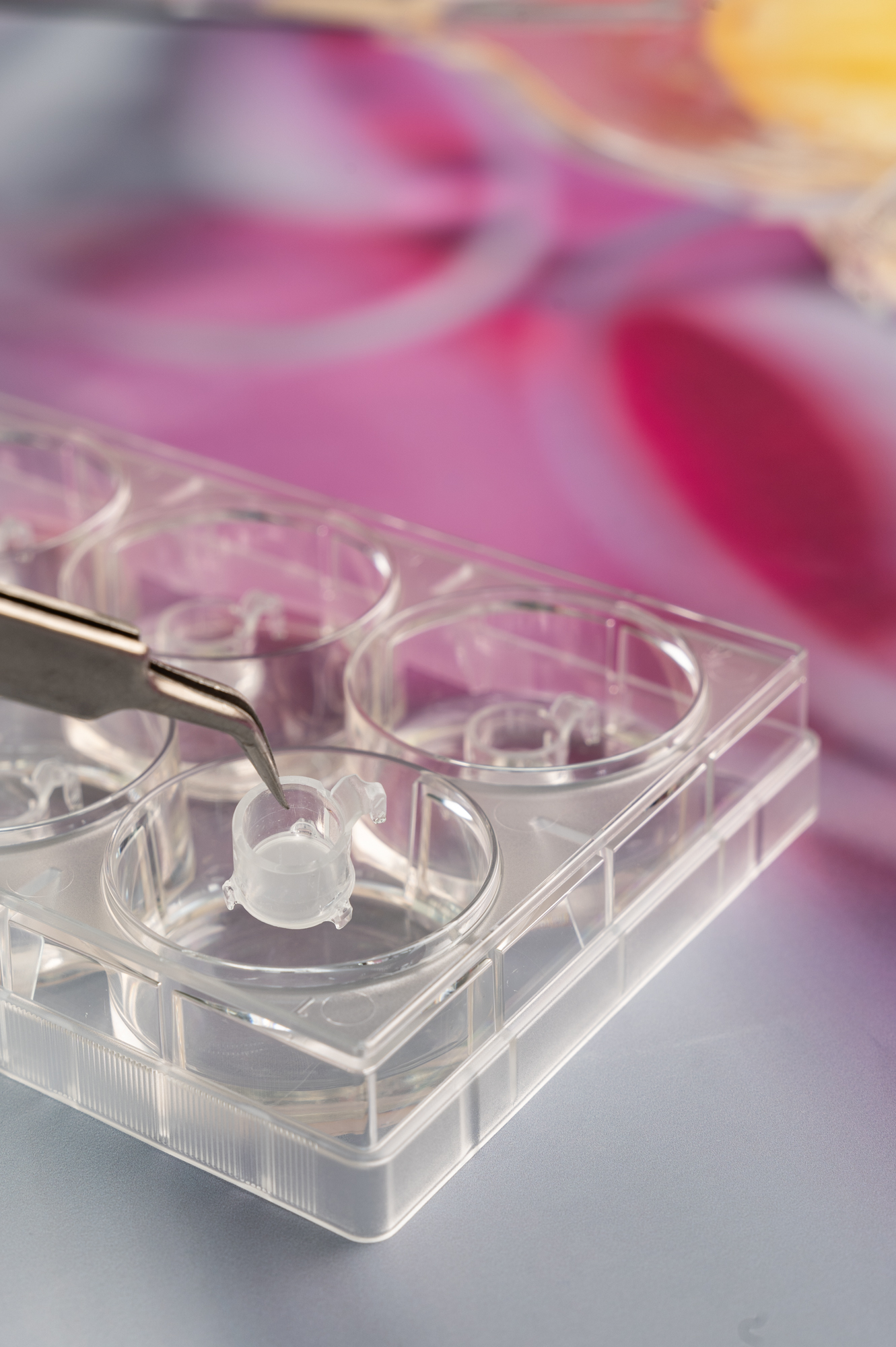
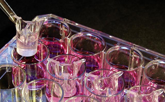
In-vitro-Hautmodell im 0,60 cm2- Format
© Fraunhofer IGB
In-vitro-Hautmodell im 0,60 cm2- Format

Our portfolio of 3D skin models 

We provide specifically suitable models for different questions and applications: 

  • Human and animal models
  • Immunocompetent skin models
  • Reporter skin models for testing substances for cell stress-influencing effects: anti-/inflammatory, sensitizing, anti-/oxidative, stress-inducing due to defective protein folding in the endoplasmic reticulum 
  • Microbiome and infectious skin models with bacteria, fungi and viruses
  • Skin disease models

Our 3D in-vitro skin models can be constructed in the following formats:

  • 4.38 cm2 (6-well)
  • 0.60 cm2 (24-well)
  • 0.38 cm2 (12-well)

Our services

We offer a wide range of services related to our skin models, which we carry out in state-of-the-art laboratories on behalf of and in collaboration with customers:

  • Conducting studies and substance screenings
  • Development of innovative and complex skin models including
    • Isolation of primary cells
    • Production of genetically modified cell lines (stable and transient)
    • Reporter cell lines with different intracellular and secreted reporters
    • 3D in-vitro tissue reconstitution
    • Phenotypic and functional characterization of models
  • Provision of all skin models for shipping

Analysis methods

The effects of chemicals, active ingredients and products on the architecture of the tissue and physiological properties can be investigated in our 3D skin models. The analyses of full skin models can be performed with the entire tissue as well as separately in dermis and epidermis. 

We offer the following analysis methods:

  • Vitality measurements, for example using the MTT test (colorimetric test), alarmaBlueTM . . .
  • Multiplex analysis for the detection and quantification of secreted proteins such as cytokines, chemokines, growth factors and antimicrobial peptides,
  • Testing of transepithelial electrical resistance (TEER) and alternative testing of the skin's barrier function,
  • Histological and immunohistochemical staining of tissue thin sections,
  • Protein analysis by Western blot,
  • Analysis of specific signaling pathway activation via reporter proteins and
  • RNA and DNA analyses using PCR or qPCR and sequencing