Printable 3D matrices for the engineering of bioartificial cartilage

The challenge of regenerating articular cartilage

Challenge: Regeneration of articular cartilage.

Because of the lack of circulation articular cartilage has no access to regenerative cell populations. Cartilage damage is therefore close to irreversible and frequently results in progressive destruction of the joint affected. One promising therapy is matrix-associated autologous chondrocyte transplantation (MACT), in which a suitable material (matrix) is seeded with the patient's cartilage cells (chondrocytes) and then implanted into the damaged cartilage. However, the cultivation of the chondrocytes of the generally used collagen-based matrices can lead to dedifferentiation, i.e. a loss of cellular function.

Reproduction of tissues by modifying natural tissue components

Reproduction of tissue by modifying natural tissue components.

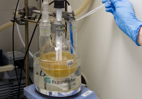
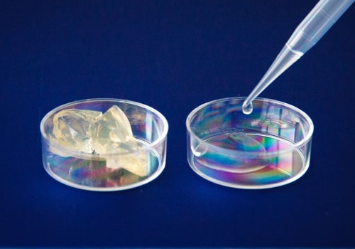
In order to preserve the function of chondrocytes it seems highly significant to create a reproduction of the native extracellular matrix (ECM) that is as natural as possible. Articular cartilage has outstanding properties in regard to strength and water content. These are due to the composition of its ECM, of collagen fibers and hydrophilic polysaccharide units (glucosaminoglycans). In order to represent cartilage-like hydrogel systems, researchers at the IGB modified biological molecules of the natural ECM by means of a chemical reaction with methacrylic acid, thereby making crosslinking possible. A two-component system made of gelatin (denatured collagen) and chondroitin sulphate (glucosaminoglycan) can thereby be chemically crosslinked into ECM-mimicking hydrogels in a controlled way.

By varying the degree of crosslinking and solid content we were able to produce gelatin hydrogels with strengths of about 5 kPa to 370 kPa. This approximately corresponds to the strength of soft fatty tissue and nasal cartilage, respectively [1]. The integration of chondroitin sulphate enables the swelling ability of the matrices to continue being increased, while retaining their strength. Thus we could improve the hydrogel properties and increase the similarity to native articular cartilage.

Stabilization of chondrocytes: the right matrix composition provides biofunctionality

Stabilization of cartilage cells.

A distinct effect of the composition of the hydrogel on the morphology and proliferative behavior of the cells was found during the encapsulation of chondrocytes in three-dimensional hybrid hydrogels. By contrast with hydrogels containing collagen or pure gelatin, chondrocytes in hydrogels containing chondroitin sulphate showed a cell type-specific spherical morphology and low cell division activity. Our biomimetic hydrogels, which imitate the natural cartilage environment, therefore represent a promising 3D system for the construction of replacement cartilage tissue.

Cell matrix systems as bio-inks to print tissues

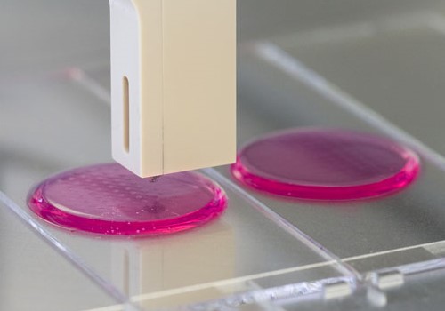
Like many other native tissues, hyaline cartilage has a characteristic micro- and macro-structure. For example, the content of proteoglycans continually increases from the joint line to the bone. Also, there are zones with high cell density as well as cell-free zones. Precise dosing techniques are necessary to be able to reconstruct the internal structures of tissues; inkjet printing is one such technique. In order to make the material systems presented here suitable for inkjet printing, the gelling characteristics of the biomolecule solution must be well-controlled prior to crosslinking and the viscosity must be kept low.

The twofold modification of the biomolecules, with crosslinking groups on one hand and with additional non-crosslinking units on the other, enables the properties of the non-crosslinked solutions and those of the crosslinked hydrogels to be adjusted independently. It is thereby possible to print chondrocytes in the gelatin-based "bio-inks" onto suitable substrates using inkjet printing [2].

Biomimetic biomaterials: a model for the future

The material systems shown here therefore have three properties that especially qualify them for constructing functional tissue models:

(1) They are based on natural extracellular matrix biomolecules.
(2) They can be adjusted to the mechanical properties of various tissues.
(3) They can be made into the desired structures using additive digital process such as 3D printing [3].

This means they have great future potential to contribute to the construction of functional tissue-replacement materials.

References

[1] Hoch, E.; Schuh, C.; Hirth, T.; Tovar, G.E.M.; Borchers, K. (2012) Stiff gelatin hydrogels can be photo-chemically synthesized from low viscous gelatin solutions using molecularly functionalized gelatin with a high degree of methacrylation, Journal of Materials Science: Materials in Medicine23:2607–2617
[2] Hoch, E.; Hirth, T.; Tovar, G.E.M.; Borchers, K. (2013) Chemical tailoring of gelatin to adjust its chemical and physical properties for functional bioprinting, Journal of Materials Chemistry B. The Royal Society of Chemistry 1: 5675-5685
[3] Engelhardt, S.; Hoch, E.; Borchers, K.; Meyer, W.; Krüger, H.; Tovar, G.;Gillner, A. (2011)Fabrication of 2D protein microstructures and 3D polymer-protein hybrid microstructures by two-photon polymerization, Biofabrication 3: 025003

Funding

We would like to thank the Max Buchner Research Foundation for funding this research.