Mammary carcinoma – malignant cell proliferation in the breast – is believed to be the most widespread form of cancer among women in Germany. In industrialized countries one in ten women will suffer from breast cancer during her lifetime. With 45,000 incidences and 18,000 mortalities per year in Germany, not only is the disease of great concern to patients but is also a significant factor in health care expenditure.
Diagnostics for individualized therapy of breast cancer
Joint research project to develop diagnostics
The onset of tumors is a multi-factorial process involving several cellular signaling pathways. Since mid 2002, scientists from the Fraunhofer IGB, together with partners at the universities of Stuttgart and Tübingen as well as the Robert Bosch Hospital, Stuttgart, have been working on a collaborative research project funded by the state of Baden-Württemberg on the molecular analysis of breast cancer tumors. The Fraunhofer IGB's role is focused on testing a DNA biochip for improved and individualized breast cancer diagnosis.
As a rule, current diagnosis procedures initially involve feeling for lumps in the breast, which are then subsequently investigated using imaging techniques such as mammography. If cancer is still suspected at this stage, the degenerate tissue is surgically removed and analyzed. It is well known that histopathologically identical tumors can exhibit different clinical progression under the same therapy.
In order to optimize therapy, there is an urgent need to further differentiate diagnosis. The creation of individual transcription profiles of tumors based on biochips can provide an appropriate approach here.
DNA biochips for advanced breast cancer diagnostics

Closely coordinating with its project partners, the Fraunhofer IGB is exploiting its long-standing expertise to develop DNA microarrays and test them for use in routine clinical diagnosis. Relevant genes are selected, tumor samples analyzed and the quality of the biochips examined. The project is also focusing on enhancing the sensitivity of the technology, so that in future it will be possible to obtain reliable analyses from smaller clinical sample amounts, such as biopsies. This includes establishing processes for RNA amplification as well as for isolation of RNA from paraffin-embedded tissues.
Design and quality control of microarrays
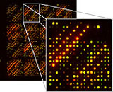
The biochip tested at the Fraunhofer IGB comprises a selective combination of several hundred genes for the classification of mammary carcinomas. A dysfunction of these genes often entails in the onset of breast carcinomas. The set serves to compile a highly informative gene transcription profile and to identify correlations with clinical processes.
This is intended to enable future use in both prognosis and to accompany therapy as part of routine clinical diagnosis. Of particular interest are the genes which regulate cellular proliferation processes. Using these genes, the array can also be deployed in general to measure proliferation in suitable cellular systems. Thus biochips are ideally suited as molecular biological tools for revealing cellular signal transduction pathways.
Cell lines for analysis of tumor samples


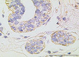
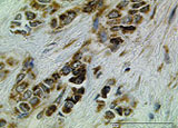
Initially, the biochip was developed using established breast cancer cell lines, which were stimulated by treatment with anti-estrogens or cytokines. Subsequently, the data generated was validated using alternative quantification methods. The biochip is used in the analysis of a large number of archived tumor preparations, stored in a tissue bank at the Robert Bosch Hospital.
Efficient data analysis
Analysis of the extensive data is carried out in collaboration with the Institute of Stochastics and Applications at the University of Stuttgart. The M-CHiPS microarray data warehouse and analysis tool developed as part of a research collaboration (German Cancer Research Center DKFZ Heidelberg, www.mchips.org) continues to be used. Thus it is possible to compile significant transcription profiles and establish correlations with clinical processes. These will facilitate the use of the biochip in routine clinical diagnosis, both for prognostic purposes and to accompany therapy.
 Fraunhofer Institute for Interfacial Engineering and Biotechnology IGB